
Available online at http://www.jo urnalcra.com
Internatio nal Journal of C urrent Research Vol. 12, Issue, 01, pp.9373-9380, Janua ry, 2020
DOI: https://doi.org/10.24941/ijcr .37766.01.2020
RESEARCH ARTICLE
INTERNATIONAL JOURNAL OF CURRENT RESEARCH
DENTAL AGE ASSESSMENT BY PULP-TO-TOOTH VOLUME RATIO OF MAXILLARY CANINE BY CERVICAL AXIAL SECTION OF CBCT- A RETROSPECTIVE STUDY OF NORTH INDIAN POPULATION
*Dr. Vikash Ranjan, Dr. Sambuddha Chakrabarty, Dr. Soumendu Bikash Maiti and Dr. V. Naveen Shankar
Department of Oral Medicine and Radiology, Divya Jyoti College of Dent al Sciences and Res earch, Modinagar
Article History:
Received 25th October, 2019 Received in revised form 18th Novembe r, 2019
Accepted 29th Decem ber , 2019 Published online 30th January , 2020
Key Words:
Age Estimation;
Cone-Beam Computed Tomogra phy ; Pulp-Tooth Volume Ratio.
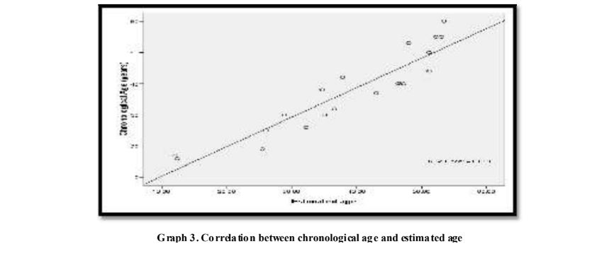
Background: Age estimati on from teeth is a key indicator to establish identity in forensi c cases. CBCT provides several advantages compared to conventional radiog raphic meth ods which has been used regularly in forensic cases. Purpos e: Dent al Age estimation by pulp-to-tooth volu me ratio using cone-beam computed tomog raphy. The Retrosp ective study will attempt to est ablishing a correlation between the chronological age of a certain individual and the pulp/tooth volume rati o using Cone- beam computed tomog raphy. Materials and Methodology: In this study a total of 50 scans (25 males and 25 females ) resp ectively were collected from the archives of a imaging cent re located in Delhi- NCR region . Out of this 50 scans, 20 scans (i.e 10 males and 10 fe males ) of age group between 15-75 years were selected for the study based on the inclusion & exclusion criteri a of the study. Resul ts: Mean difference and Standard error difference bet ween the Chronological and Esti mated age was not si gnificant as p> 0.05 , that is the difference bet ween the Chronologi cal and Estimated age was less. The Pearson Correlation (r) were highly significan t, p<0.01. Esti mated age was more accurate in mid dle age. Regression formula for maxil lary canine (overall) Age = 48.009 – (973 .172 × Pulp/ Tooth Volume Ratio). Concl usion: In our study stand ard error difference between chronological and estimated age is ±4.09781 years. There was a negative correlation between chronological age and Pulp/ tooth volume ratio, as the advancing age is asso ciat ed with a decrease in the pulp/ tooth volu me ratio.
Copyright © 2019, Vikash Ranjan et al. This is an open access article distributed under the Creative Commons Attribution Lice nse, which permits unrestricted use, distribution, and reproduction in any medium, provided the original work is properly cited.
INTRODUCTION
Age estimation is one of several indicators employed to establish identity in fo rensic cases. Most frequ ently, Age estimations from t eeth are us ed, as teeth may be ret ained long after all other tissues, even bone, have d egenerated, but unlike bone they can also be investigated directly in living individuals. The dental age estimation methods most o ften requires extraction, and some of them also need prepared for microscopic s ections o f at least one tooth. T hese methods are tedious and costly, and a damaging approach may not be acceptable for ethical, r eligious, cultural, or scienti fic reasons. A study of radiographs of the teeth is a non-destructive, straightfo rward method to obtain in formation and is a technique used regularly in most dental surgeries, but it is rarely employed in methods of age estimation (Kvaal, 1995). Techniques for chronological age estimation in children b ased on dental maturation may b e divided into those using the atlas approach and those using scoring system whereas in adults there are morphological and radiog raphical techniques (Willems, 2001). Changes that are detectable with increasing age are attrition, periodontal disease, and deposition of secondary dentine, root t ranslucency, cementum apposition, root resorption, color changes and increase in root roughn ess. Gustafson in 1950 suggested the use of six retrogressive changes and ranked them on arbitrary scale, allotting 0-3 points according to degree of the change. Error obtained in this morphometric method resulted in several modification in subsequent studies. Johanson in1971 in his research used same six criterions but different ranking scale and then estimated the age of an individual. Solheim used in situ teeth and eight variables which included two of color estimate, two for periodontosis, and two for attrition, crown length and sex (Singh, 2004). The apposition of secondary dentine is also oft en taken into account, because the pulp is surrounded not only by hard tissue such as enamel but also by dentine, which changes du ring an individual’ s life. Canines were chosen for a number of reasons: they have the longest fun ctional survival rate in the mouth, undergo less wear as a result of diet than posterior teeth, and also su ffer less wear than other anterior teeth due to th eir particular work, and have the largest pulp area amongst the single rooted t eeth and thus the easiest to analyze (Cameriere, 2009). CBCT provides a non-invasive alternative for age estimation which is an important aspect of fo rensic dentistry. Enough data for volumetric image construction is captured by a single rotational sequence. The target region is scanned in a single rotation there fore, the radiation exposure is less. CBCT may be very useful in some fo rensic p rocedures, o ffering several advantages fo r pre- mortem forensic and post-mortem forensic imaging including good resolution fo r skelet al imaging, rel atively low cost, portability, and simplicity. The pulpo-dentinal complex (dentin, cementum, and the dental pulp) shows physiologic and pathological changes with advancing age (Jawaid, 2014).
MATERIALS AND METHODS
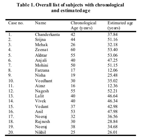
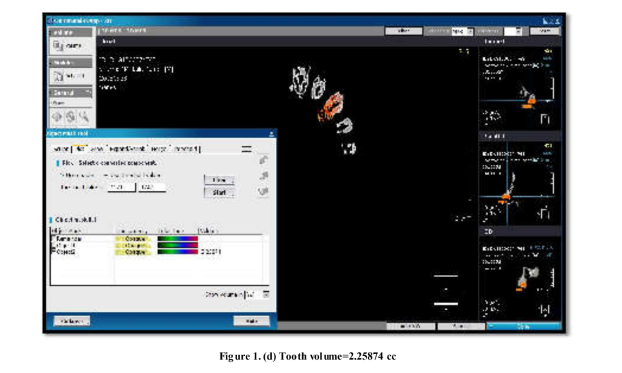
This retrospective double blind study comprised a total of 50 CBCT scans i.e 25 males and 25 females resp ectively were collected from the archives of a imaging cent re located in Delhi-NCR region. Out of this 50 CBCT scans, 20 CBCT scans (i.e 10 males and 10 femal es) o f age group between 15- 75 years were s elected fo r the study based on the inclusion & exclusion criteria o f the study. Inclusion Criteria comprising of patients within the age range of 15-75 years & canine upto root apices. Exclusion Criteria are canine with caries, filling, or crown restoration, periapical pathologies, anomalies, fracture of maxilla & space in fection. CBCT scans were obtained for various reasons such as impacted teeth, dental anomalies, implant planning or orthodontics. Single maxillary canine tooth were assess ed retrosp ectively from patients. Teeth with caries, filling or crown restorations, periapical pathologies anomalies or pulpal pathosis were excluded. All CBCT images were taken with the sam e device ( Kodak CS9300, Carestream Health, Rochester, NY) using 60–90 kVp, 2–15 mA and 12–28 s scanning time. For volumetric measurements, CBCT scans were exported as DICOM files and then imported into a volumetric rendering so ftware capable of measu rements of vector-based segmentation in On Demand software. On each of CBCT image maxillary canine was sel ected. Imag e of axial section at the cemento-enamel junction was obtained. Pro file tool was selected for getting minimum and maximum threshold of pulp of each individual. After Segmentation of tooth and pulp cavity, then pick tool was used in which minimum and maximum threshold had been entered, then desired area such as tooth and pulp was marked with the pick tool, separately using their respected maximum and minimum threshold, then the volume was calculated in cc. Aft er all measurements, pulp volume to tooth volume ratio was calcul ated. All measurem ents were performed by a observer no.1 without any knowledge of chronological age. After the study, Estimated age will be compared with Chronological age in the presence of observer no. 2.
RESULTS AND DISCUSSION
Several studies suggested that the measurement of the pulpal size area in canines revealed better secondary dentinal deposition than the other teeth groups with smaller pulp areas. Smaller size o f such single-rooted teeth may lead to less clear images and inaccurate pulp tooth ratio measurement
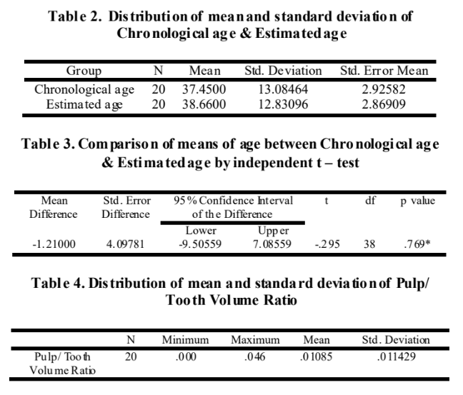
Therefore, in the present study we did not include central and lateral incisors owing to their small pulpal sizes. In the present study there was a highly significant correlation R= -0.850 between chronological age and Pulp/ tooth volume ratio in the given set o f data and the study conducted by Jagannath an N et al. (2011) which showed a similar correlation R = -0.63. In the study by Jagannathan et al. (2011), in the study group produced an MAE o f 15. 34 years in 72.81% of the cases. Age estimates were within ±10 years o f actual age in 27.09% of the cases. Application of the formula derived by Jagannathan et al. (2011) to the control group yielded an MAE of 8.54 years whereas in our study standard error diference between chronological and estimated age is 4.09781 years. In a study by Somedaetal. (2009) females tended to have higher accuracy compared to males, which is contrary to our study where males tended to have higher accuracy compared to females as standard error mean fo r males (3.89556 years) is less than females (4.38869 years). The R2 value indicates how much o f the dependent variable (Chronological age) can be explained by the Independent variable (Pulp/tooth volume ratio). In our study, R2= 72.3% which is a significant which is a significant value than the result given by study of Yang F et al6 that is R2=29%.
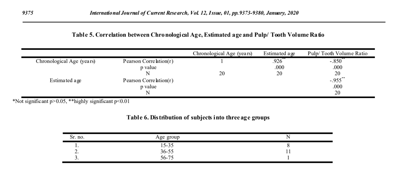
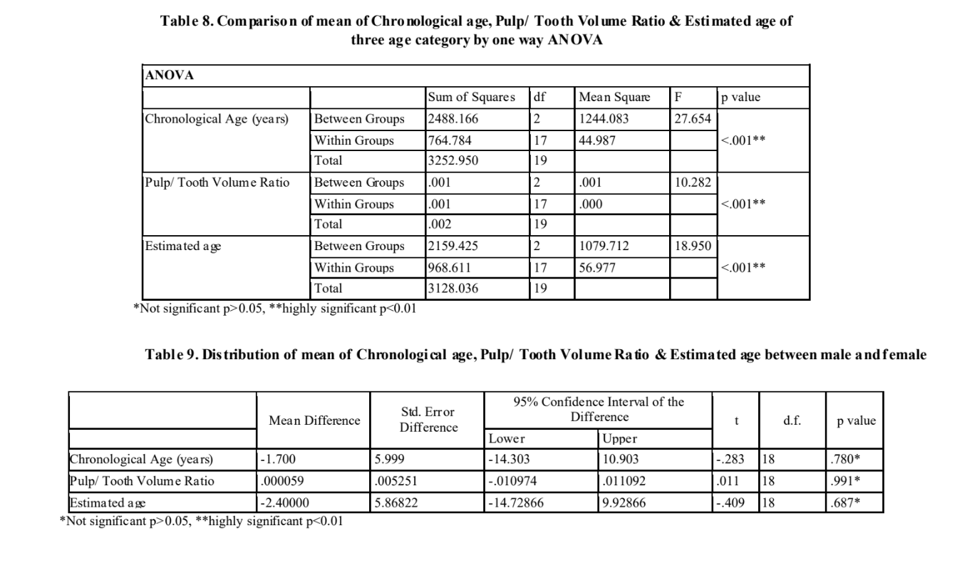

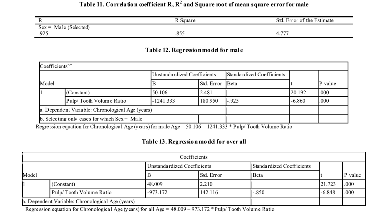
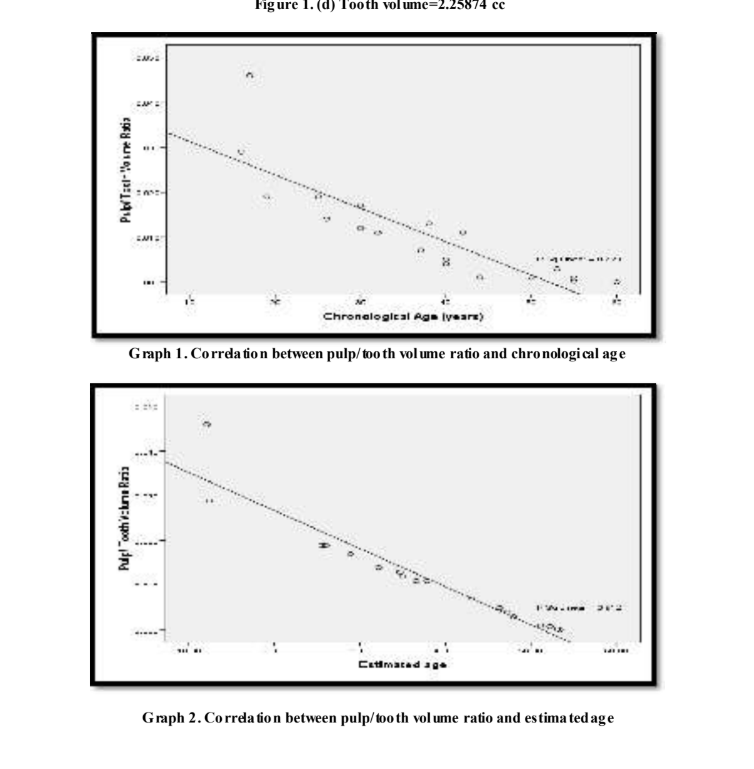
The use of Yang’s formula in the control group produced an estimated age has a standard deviation ± 14.78 years, whereas in our study an estimated age has a standard error difference ±4.09781 years. The square root of mean square error was 7.080 years where it was 8.3 years in the study by Yang F et al6. In the study by Yang F et al6, The correlation between age and Pulp/Tooth Volume ratio is -0.54 whereas in our study correlation between age and Pulp/ Tooth Volume Ratio is – 0.850, which is highly significant, p<0.01.This states that Pulp/Tooth Volume Ratio is inversely p roportional to age. There was a negative (inversely proportional) correlation between chronological age and Pulp/ tooth volume ratio, as the advancing age is associated with a decrease in the pulp/ tooth volume ratio as mentioned by Yang et al, Jagannathan et al. (2011) Gulsahi et al. (2017) Babshet et al. (2010) performed a study on Indian sample, resulting in an MAE of 10.76 years whereas in our study standard error di fference between chronological and estimated age is 4.09781 years. Also, In a sudy by Ge et al. (2015) the mean absolute error was 8.122whereas in our study standard error di fference between chronological and estimated age is 4.09781 years. In a study by Gulsahi et al. (2017) there was no signi ficant di fference in the intercept b etween both gender (p > 0.05). This study revealed that PV/TV ratio was not gender dependent. In the present study, the mean differen ce between male and female of Chronological Age is -1.700 and p value is 0.780, of Pulp/ Tooth Volume Ratio is 0.00059 and p value is 0.991 and of Estimated age is -2.400 and p value is 0.687, which are not significant, p>0.05. In a study by Rangari et al. (2018) there was a moderately signi ficant correlation i.e. R = -0.599 between chronological age and Pulp/ tooth volume ratio in the given set of data for all teeth, R= -0.533 fo r maxillary central incisor and R=0.562 for maxillary canine. In the present study, there is signi ficantly high correlation i.e. Correlation( r) between Chronological Age and Pulp/ Tooth Volume Ratio R= -0.850. Study conducted by Rangari et al. (2018) determined R2 = 35.8% for all teeth, R2 = 31.6% for maxillary canine and R2= 28.4% can be explained for maxillary central incisor, which were moderately signifi cant whereas the present study determined, R2 =72.3% for maxillary canine whi ch is highly significant. The square root of mean square error was 11.45 years whereas in the present study, Square root of mean square error is 7.080 years. V arious studies have obtained regression fo rmula for calculating age by using pulp/tooth volume ratio such as Yang et al. (2006) obtained the equation of the straight line relating age and ratio of pulp/tooth volume estimated as: Age = 54. 32 – (554.21 ×pulp/ tooth volume Ratio). Jagannathan et al. (2011) obtained the regression equation for the Indian population for maxillary canine: Age = 57.18 + (- 413.41 x pulp/tooth volume ratio). Rangari et al. (2018) obtained the Regression formula fo r maxillary canine as Age= 53.418+(-1415.733×PTVR). Gulsahi et al. (2017) obtained the Regression formula for maxillary canine as Age = 60.5 − (479.3 × PV/T V). In our study, Regression analysis yielded a statistically significant but moderate negative correlation between pulp/tooth volume ratio and age. Regression formula fo r maxillary canine for m ales is Age = 50.106 – (1241.333 × Pulp/ Tooth Volume Ratio) and for femal es is Age = 47. 695 – (868.211 × Pulp/ Tooth Volume Ratio). Regression formula fo r maxillary canine (ov erall) Ag e = 48.009 – (973.172 × Pulp/ Tooth Volume Ratio).
Conclusion
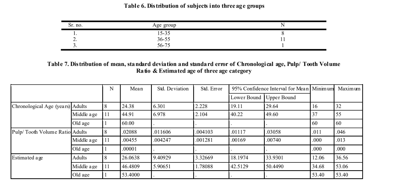
Mean difference between Chronological age and Estimated age is not significant as the difference between the Chronological and Estimated age is less. The Pearson Correlation(r) between Chronological Age and Estimated age, Chronological Age and Pulp/ Tooth Volume Ratio, Estimated age and Pulp/ Tooth Volume Ratio are highly signi ficant, p<0.01. Estimated age is more accurate in middle age (36-55 years) than young adults (15-35years) and old age (56-75). Regression formula for maxillary canine for males is Age = 50.106 – (1241.333 × Pulp/ Tooth Volume Ratio) and for femal es is Age = 47. 695 – (868.211 × Pulp/ Tooth Volume Ratio). Regression formula fo r maxillary canine (ov erall) Ag e = 48.009 – (973.172 × Pulp/ Tooth Volume Ratio).
The presented method is a promising tool in the procedure for age estimation, permitted by the high technological level achieved by the currently available machines fo r the CBCT. Further advancements could help optimizing the accuracy and precision of the technique. Recent generations in cone-beam CT have become available, demonstrating better contrast resolution. T he CBCT may bring more detail in the grayscale level rang e and enable improved visualization of the tooth segmentations. A large data sample with homogeneous (or equal) age distribution should allow for even more finesse and optimization of the elaborated method. Also a large data sample on large geographical area is required for more accuracy o f the result. T hat would allow forensic odontologist to use the present method for age estimation using a very objective technique.
REFERENCES
Kvaal SI., Kollviet KM., Thomsen O., Solheim T. 1995. Age estimation of adults from dental radiographs. Forensic Science International., 1995;74:175-185.
Willems G. 2001. A Review of the most commonly used dental age estimation techniques. The Journal of Forensic Odonto-Stomatology., June19:1.
Singh A., Gorea RK., Singla U. 2004. Age estimation from the physiological changes of t eeth. Jiafm.; 26(3):0971- 0973.
Cameriere, R., Ferrante L., Cingolani M. 2009. Age estimation by pulp/tooth area ratio in canines: Study of a Portuguese sample to test Cameriere’s method. Forensic Science International.193:128.e1–128.e6.
Jawaid M, Iqubal MD, Shukla AK, Khan M, Farh B. 2014. The role of cbct in forensic dentistry: a review. International Journal Of Advances In Case Reports., 1(4):179-183.
Yang F, Jacobs R, Willems G. Dental age estimation through volume matching of teeth imaged by cone-beam CT. Forensic Science International. 2006;159:78–83.
Someda H., Matsunaga S. , Ide Y., Nakahara K., Hirata S. , Hashimoto M. 2009. Age estimation based on three- dimensional measurement o f mandibular central incisors in Japanese. Forensic Science International., 185:110–114.
Babshet M., Acharya AB., Naikmasur VG. 2010. Age estimation in Indians from pulp/tooth area ratio of mandibular canines. Forensic Science International., 197:125.e1–125.e4.
Jagannathan N., Neelak antan P. , Thiruvengadam C., Ramani
- , Premkumar P., Natesan A., Herald JS., Luder HU. 2011. Age estimation in an Indian population using pulp/tooth volume ratio of mandibular canines obtained from cone beam computed tomography. J Forensic Odontostomatol., 29:1:1-6.
Ge ZP., Ma RH., Li G., Zhang JZ., Ma XC. 2015. Age
estimation based on pulp chamber volume of first molars from cone-beam computed tomography images. Forensic Science International., 253:133.e1–133.e7.
Gulsahi A, Kulah CK, Bakirarar B, Gulen O, Kamburoglu K. 2017. Age estimation based on pulp/tooth volume ratio measured on cone-beam CT images. Dentoma xillofacial Radiology. 46:239.
Rangari P. , Singh N., Cheema B., Singh U. 2018. Age Estimation Using Pulp/ Tooth Volume Ratio By Cone Beam Computed Tomography. OSR Journal of Dental and Medical Sciences., 17(3):18-23.
*******